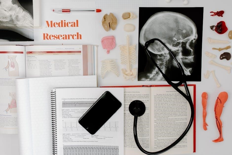
The human anatomy lab manual offers a comprehensive guide for exploring anatomy through cat dissections.
- Provides hands-on learning opportunities.
- Covers all body systems.
- Includes clear instructions and visual aids.
1.1 Overview of the Lab Manual
The lab manual provides a structured approach to studying human anatomy through cat dissections‚ offering a detailed exploration of all body systems. It includes 30 exercises designed for a one-semester course‚ ensuring a comprehensive understanding of anatomical structures. Full-color illustrations and clear instructions guide students through dissection procedures‚ while clinical application questions enhance critical thinking. The manual is tailored for hands-on learning‚ making it an essential resource for anatomy students. Its engaging writing style and visual aids ensure an effective learning experience‚ preparing students for successful lab work and practical applications in the field of anatomy.
- Covers all body systems in depth.
- Includes 30 exercises for practical learning.
- Features full-color illustrations for clarity.
- Encourages critical thinking with clinical questions.
1.2 Importance of Cat Dissections in Anatomy Study
Cat dissections are a valuable tool in anatomy education‚ offering a hands-on approach to understanding anatomical structures. Cats possess anatomical similarities to humans‚ making them an ideal model for studying muscle‚ skeletal‚ and organ systems. Dissection allows students to visualize and explore complex structures in a three-dimensional context‚ enhancing their comprehension of human anatomy; It also fosters critical thinking and observational skills‚ which are essential for future healthcare professionals. By translating cat anatomy to human relevance‚ students gain practical insights into anatomical relationships and functional processes.
- Provides a hands-on learning experience.
- Enhances understanding of human anatomy through comparison.
- Develops critical thinking and observational skills.

Preparation for Cat Dissection
Proper preparation ensures a safe and effective dissection experience. Gather essential tools‚ wear protective gear‚ and review the lab manual for clear instructions.
- Ease of learning through structured guidance.
- Ensures safety and efficiency during the process.
2;1 Essential Tools and Materials Needed
Dissection requires specific tools to ensure efficiency and safety. Essential items include scalpels‚ forceps‚ tweezers‚ gloves‚ and protective eyewear. A dissection tray or pan is necessary for organizing the specimen. Magnifying glasses or dissecting microscopes may be used for detailed observations. Lab manuals provide step-by-step guidance‚ while cameras can document findings. Optional tools like hand lenses or additional lighting can enhance visibility. Ensure all materials are sterilized and within reach to maintain a smooth workflow during the procedure.
- Scalpels and blades for precise cuts.
- Forceps and tweezers for tissue handling.
- Gloves and goggles for personal protection.
2.2 Safety Precautions and Lab Etiquette
Safety and proper etiquette are crucial during cat dissections. Always wear gloves‚ goggles‚ and a lab coat to protect against biohazards. Handle sharp instruments like scalpels with care‚ ensuring proper technique to avoid injuries. Dispose of biological waste and materials according to lab guidelines. Maintain a clean workspace to prevent contamination. Respect shared equipment and follow instructions provided by instructors. Avoid eating or drinking in the lab. Collaborate with lab partners and ensure all actions align with ethical practices. Familiarize yourself with emergency procedures‚ such as eye wash stations and first aid kits‚ in case of accidents.
- Wear protective gear at all times.
- Handle sharp tools with caution.
- Follow proper waste disposal protocols.
Skeletal System Exploration
The skeletal system exploration involves identifying and studying cat bones and joints through dissection. This provides insights into human skeletal structures by comparison‚ aiding anatomy understanding. Key bones like the skull‚ vertebrae‚ and limb bones are examined to understand their roles and relationships. Visual aids and detailed descriptions guide students in recognizing anatomical features‚ fostering a deeper appreciation of skeletal functions and their relevance to human anatomy.
3.1 Identifying Key Bones and Joints
In this section‚ students learn to identify and examine the primary bones and joints of the cat skeleton. Using detailed diagrams and bone charts‚ participants compare feline anatomy to human skeletal structures. Key bones such as the femur‚ humerus‚ and vertebrae are highlighted‚ along with their functions and connections. Joints‚ including ball-and-socket and hinge types‚ are explored to understand their roles in movement and support. Hands-on activities involve palpation and dissection to locate and label bones and joints‚ fostering a deeper understanding of skeletal anatomy and its relevance to human physiology.
3.2 Dissection of the Skull and Vertebrae
Dissection of the skull and vertebrae provides a detailed exploration of the cat’s axial skeleton. Students carefully expose the skull‚ identifying its structure‚ including the cranium‚ facial bones‚ and foramen. The process involves removing the calvaria to observe the brain and its protective membranes. Vertebrae are examined to understand their classification (cervical‚ thoracic‚ lumbar) and functions‚ such as spinal cord protection and nerve root exits. This exercise enhances understanding of skeletal anatomy‚ offering insights into human anatomical parallels. Hands-on activities include labeling bones and joints‚ fostering familiarity with complex structures and their roles in movement and support.

Muscular System Analysis
This section focuses on skin preparation and muscle identification through detailed dissections of forelimb and hindlimb muscles‚ offering insights into their structure and function.
4.1 Skin Preparation and Muscle Identification
This section guides students through preparing the cat specimen for muscular system analysis. Proper skin removal techniques are emphasized to expose underlying muscles. Tools like scalpels and forceps are used to carefully retract tissue‚ ensuring muscles remain intact. Detailed instructions help identify major muscle groups‚ focusing on their attachments‚ actions‚ and relationships with other structures. High-quality illustrations and descriptions aid in distinguishing between superficial and deep muscles. Students learn to recognize the forelimb and hindlimb muscles‚ laying a foundation for understanding human anatomy through comparative study. This hands-on approach enhances anatomical knowledge and develops dissection skills essential for future clinical applications.
4.2 Detailed Dissection of Forelimb and Hindlimb Muscles
This section provides a step-by-step guide for dissecting the forelimb and hindlimb muscles of the cat specimen. Students learn to identify and isolate major muscle groups‚ such as the flexors‚ extensors‚ and rotators‚ using precise dissection techniques. The process involves careful removal of connective tissue to expose muscle origins and insertions. Comparisons are drawn between feline and human musculature‚ highlighting functional similarities. Detailed illustrations and descriptions aid in recognizing muscle layers and their relationships. This exercise enhances understanding of muscle anatomy and their roles in movement‚ preparing students for clinical applications in human anatomy and physiology.
Respiratory and Digestive Systems
This section focuses on the examination of the respiratory and digestive systems through cat dissections. Students explore the thoracic cavity‚ lungs‚ and abdominal organs in detail.
5.1 Examining the Thoracic Cavity and Lungs
Examining the thoracic cavity and lungs is a critical part of understanding respiratory anatomy. Students learn to carefully dissect and expose the ribcage‚ diaphragm‚ and lungs. Key structures such as the trachea‚ bronchi‚ and pulmonary vessels are identified. The process involves removing the sternum and ribs to access the thoracic organs. Observing the lung lobes and their relationship to the diaphragm provides insights into respiratory mechanics. This exercise helps students visualize how the lungs expand and contract during breathing. The detailed dissection enhances comprehension of human respiratory anatomy through comparative cat dissection.
5.2 Dissection of the Abdominal Cavity and Organs
Dissecting the abdominal cavity provides in-depth insights into the arrangement and function of digestive organs. Students carefully remove the abdominal wall to expose the peritoneal cavity. Key organs such as the stomach‚ liver‚ pancreas‚ spleen‚ and intestines are identified. The process involves tracing blood vessels and nerves supplying these organs. Observing the structural relationships between organs‚ like the liver’s position under the diaphragm‚ enhances understanding of digestive system anatomy. This hands-on exercise helps students correlate cat anatomy with human digestive physiology‚ emphasizing clinical relevance and functional anatomy.

Circulatory and Nervous Systems
This section explores the cat’s circulatory and nervous systems‚ focusing on heart structure‚ blood vessel mapping‚ and brain dissection‚ revealing their interconnected roles in human anatomy.
6.1 Mapping Blood Vessels and the Heart
This section focuses on identifying and mapping the major blood vessels and examining the structure of the cat’s heart. Students learn to trace the arterial and venous pathways‚ understanding their roles in circulation. The heart is dissected to reveal its chambers‚ valves‚ and coronary vessels‚ providing insights into cardiac anatomy. This exercise helps correlate cat anatomy with human cardiovascular systems‚ essential for future healthcare professionals. Key procedures include identifying the aorta‚ pulmonary arteries‚ and renal veins‚ as well as exploring the heart’s internal structures. This hands-on approach enhances understanding of blood flow dynamics and their clinical relevance in human health.
6.2 Exploring the Brain and Spinal Cord
This section involves the detailed dissection and examination of the cat’s central nervous system‚ focusing on the brain and spinal cord. Students identify key structures such as the cerebrum‚ cerebellum‚ and brainstem‚ as well as the meninges and cranial nerves. The spinal cord is explored to understand its segmentation and relationship with the vertebral column. This exercise provides a foundation for understanding human neuroanatomy‚ as the cat’s nervous system shares similarities with humans. The dissection highlights neural pathways and the protective mechanisms of the central nervous system‚ offering practical insights into the functional anatomy of the brain and spinal cord.

Clinical Applications of Cat Dissection
Cat dissection aids in understanding human diseases‚ surgical techniques‚ and diagnostic methods. It bridges anatomical knowledge to real-world medical practices‚ enhancing skills in patient care and treatment.
7.1 Translating Cat Anatomy to Human Anatomy
Cat anatomy shares significant similarities with human anatomy‚ making it an effective model for studying human structures and systems. For instance‚ the feline skeletal and muscular systems closely resemble those of humans‚ allowing for accurate comparisons. The arrangement of organs in the thoracic and abdominal cavities also mirrors human anatomy‚ facilitating the study of organ function and disease processes. Understanding these anatomical parallels enables students to apply their knowledge to human clinical scenarios‚ such as surgical procedures and diagnostic techniques. This translational approach enhances comprehension of human anatomy and prepares students for real-world medical applications.
7.2 Case Studies and Practical Implications
Cat dissection case studies provide practical insights into human anatomy and its clinical relevance. By examining feline anatomical structures‚ students can better understand human conditions‚ such as injuries‚ diseases‚ and surgical interventions. For instance‚ studying the feline cardiovascular system helps in grasping human heart anatomy and its related pathologies. Practical implications include improved surgical techniques and diagnostic skills‚ as the anatomical knowledge gained can be directly applied to human medicine; These case studies bridge the gap between theoretical learning and real-world applications‚ preparing students for clinical scenarios and enhancing their problem-solving abilities in medical contexts.
Review and Assessment
This section summarizes key concepts and provides guidelines for evaluating student understanding through quizzes‚ exams‚ and lab reports‚ ensuring comprehensive grasp of anatomical knowledge and practical skills.
8.1 Key Concepts and Takeaways
This section summarizes the essential lessons learned throughout the lab manual‚ emphasizing the importance of understanding anatomical structures and their functions. Key concepts include the identification of bones‚ muscles‚ and organs‚ as well as the interconnections between body systems. Students will gain practical skills in dissection techniques and develop critical thinking through clinical application questions. The takeaways highlight the correlation between cat anatomy and human anatomy‚ reinforcing foundational knowledge for future studies in medicine or biology. By mastering these concepts‚ students will be well-prepared for advanced coursework and real-world applications in healthcare.
- Comprehensive understanding of anatomical structures.
- Practical dissection and identification skills.
- Integration of anatomy with clinical practices.
8.2 Lab Report Guidelines and Grading Criteria
Lab reports are a critical component of the anatomy course‚ requiring detailed documentation of dissection observations and analyses. Reports must follow a structured format‚ including an introduction‚ materials‚ procedures‚ results‚ and conclusions. Grading emphasizes accuracy‚ completeness‚ and clarity in identifying anatomical structures and explaining physiological concepts. Clinical application questions are also evaluated for critical thinking and relevance to human anatomy. Proper terminology and formatting are essential‚ with penalties for errors. Submission deadlines are strictly enforced‚ and late reports may incur grade reductions. Refer to the lab manual for specific guidelines to ensure compliance with expectations and achieve optimal scores.
- Adhere to the specified report format.
- Demonstrate accurate anatomical knowledge.
- Submit on time to avoid penalties.